
Latest Research
- 2015.06.05
Molecular orientation imaging of the human hair α-keratins by VSFG-detected IR super-resolution microscope
1. Introduction
Everyone knows that microscopes are very important tools for studying biological samples. However, most of the microscopes cannot measure the vibrational molecular imaging based on molecular information. At the moment, only two microscopes are measureable. One is the Raman microscope, and the other is IR microscope, those are complementary relations. But these are not perfect. Especially, the IR microscope has very low spatial resolution about 10 µm, because of the diffraction limit by the IR wavelength. This is a serious problem for a microscope.
To solve this problem, we have developed IR super-resolution microscope with a sub-micrometer spatial resolution by using vibrational sum-frequency generation (VSFG) detection, and have reported the applications to various biological samples including living cells [1, 2]. In this study, we applied an IR super-resolution microscope to human hair samples.
2. Principle of IR super-resolution imaging
Fig. 1(a) shows a transmission image of the cross section of a human hair. The medulla at the center, the cuticle located at the rim, and the cortex in between them are distinguished in the image. In the cortex region, local fine structure can also be seen, although no molecular information is available. The conventional IR image of the sample was also measured by monitoring 3330 cm-1 absorption, which is shown in Fig. 1(b). In the conventional IR image, the medulla, the cortex and the cuticle can be distinguished; however, no further information is available because of a lack of spatial resolution. This was clearly caused by the limitation of the conventional IR microscope due to the diffraction limit.Fig. 1(c) presents a VSFG-detected IR super-resolution image of the same sample. The IR laser frequency was fixed to the NH stretching vibration at 3330 cm-1, while the visible laser wavelength was tuned to 610 nm. The observed IR super-resolution image (Fig. 1(c)) is largely different from that of the conventional IR image given in Fig. 1(b). In the conventional IR image, the cortex region is almost flat, and no fine structure can be seen. On the other hand, the IR super-resolution image consists of fine structures. This directly proves the advantage of combining the VSFG (Fig. 1(d)) and the microscope. More qualitatively, the spatial resolution is estimated to be about 0.84 µm by convolving the Gaussian point-spread-function with a step function. The resolution of 0.84 mm is roughly one order of magnitude higher than the diffraction limit of the IR light, which is 4.6 µm at this wavelength with an objective lens of NA = 0.4. Thus, the VSFG-detected IR microscope is a super-resolution one.
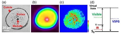 Fig. 1
(a) Optical image of a human hair cross section.
(b) Conventional IR image of the band intensity at 3330 cm−1.
(c) IR super-resolution image obtained by the simultaneous introduction of visible and IR beams (vVIS = 610 nm, vIR= 3330 cm-1). The absorbance was indicated by color from blue to red. The white scale bars in figures (a)-(c) indicate 20 µm.
(d) Schematic diagram of the VSFG process.
3. Molecular orientation imaging
Let us change the vibrational mode from NH stretching to amide vibrations in the 6-9 mm region. Amide vibrations are the most important vibrations that characterize the secondary structure of proteins. The α-helix structure of keratin protein (α-keratin) in a hair was concluded from the frequency of the amide I~III bands [3, 4].
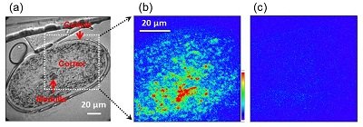
Fig. 2
(a) Transmission image of the cross section of a human hair.
(b) IR super-resolution image at the amide III vibration (vVIS = 610 nm, vIR = 1250 cm-1).
(c) IR super-resolution image at the amide I vibration (vVIS = 610 nm, vIR = 1650 cm-1).
For the amide III band (1250 cm-1), the cross-section sample gave clear strong VSFG signals at the cortex area (Fig. 2(b)). This enabled us to measure the distribution of α-keratins in the hair. On the other hand, the VSFG signal disappeared completely when the amide I band (1650 cm-1) was monitored by the same polarization of incident light (Fig. 2(c)). Why did the drastic contrast appear? Since amide I is a characteristic vibration of the a-helix as well as amide III, we expected a clear image for amide I band.
This can be explained by the molecular orientation of the α-keratin. Fig. 3 shows the molecular structure of the α-keratin with the amide I and III vibrations. Amide I mainly contributes to the CO stretching vibration, and amide III is the CN stretching vibration (Fig. 3(a)). In the α-keratin, the transition dipole of the amide I lies along the α-keratin direction, and amide III vibrates perpendicularly to it (Fig. 3(b)). In our experiment using the cross-section sample, only amide III was clearly observed. This result directly proves that the α-keratin is aligned to the longitudinal direction nearly perfectly because the cross-section sample was prepared to be perpendicular to the longitudinal direction of human hair [5].
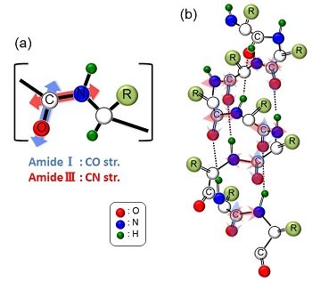
Fig. 3
(a) Vibrations of the amide I and III bands.
(b) Vibrational directions of amide I and III in the a-helix structure.
To obtain the direct evidence of our conclusion, we also measured the oblique section (α = 45 degree) of human hair. The results are shown in the Fig. 4. When a cutting angle was increased to the 45 degree, VSFG images were clearly observed even in the amide I band at 1650 cm-1. This is concluded that the molecular orientation of α- keratins was observed.
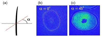
Fig. 4
(a) Cutting angle of the oblique section α.
(b) IR super-resolution image of oblique section α = 0 degree at the amide I vibration.
(c) IR super-resolution image of oblique section α = 45 degree.
4. Conclusion
We applied VSFG-detected IR super-resolution microscope to the cross section of a human hair, and attempted IR imaging at IR super-resolution in the 6-9 lm mid-IR region. In the amide III band, we clearly observed strong signals at the cortex area. On the other hand, the lack of the VSFG signal in the amide I band leads to the conclusion that the α-keratins in human hair is strongly aligned to the longitudinal direction. The results suggest that the VSFG-detected IR super-resolution microscope is a powerful tool for understanding not only molecular vibrations, but also molecular orientations and distributions in biological samples at sub-micrometer resolution.
References
[1] K. Inoue, M. Fujii, M. Sakai, Appl. Spectrosc. 64 (2010) 275.
[2] M. Sakai, K. Inoue, M. Fujii, Curr. Pharm. Biotechno. 14 (2013) 159.
[3] P. Dumas, L. Miller, Vib. Spectrosc. 32 (2003) 3.
[4] K.L.A. Chan, S.G. Kazarian, A. Mavraki, D.R. Williams, Appl. Spectrosc. 59 (2005) 149.
[5] M. Sakai, K. Kikuchi, M. Fujii, Chem. Phys. 419, (2013) 261.