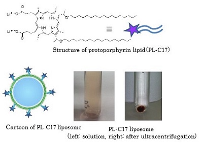
Figure 2. Structure of the protoporphyrin lipid (PL)
and its liposomal formation
PL-C17 liposomes were prepared from distearoylphosphatidylcholine (DSPC), cholesterol and distearoylphosphatidylethanolamine (DSPE)-PEG2000 (1:1:0.11, molar ratio) by the reverse-phase evapolation method. The PL-C17 micelle solution was added to the liposome solution. Figure 2 shows the liposome solution mixed with the PL-C17 micelles. After ultracentrifugation, the supernatant obtained was colorless, revealing that all PL-C17 lipids were post-inserted into the liposomal bilayer.
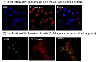
Next, we examined the localization of PL-C17 liposomes in HeLa cells. The cells were incubated with PL-C17 liposomes at 100 µM concentration for 3 h. Fluorescence images of PL-C17 liposomes in HeLa cells are shown in Figure 3A. The liposomes were localized in the Golgi apparatus in cells. Therefore, we examined that the Golgi apparatus by visualization with a green-fluorescence probe (Golgi-ID), to determine whether PL-C17 liposomes were localized there or not. Figure 3B shows the fluorescence images of PL-C17 liposomes (red) and Golgi apparatus (green) in HeLa cells. The merged image yielded orange fluorescence, thereby revealing that the PL liposomes are accumulated in the Golgi apparatus..
Figure 3. Localization of PL liposomes in cells
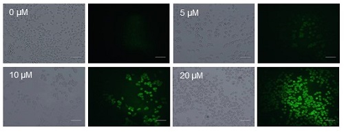
The antitumor effect of PL-C17 liposomes on HeLa cells under light irradiation was examined. HeLa cells were incubated for 3 h in medium containing various concentrations of PL-C17 liposomes. Then, the cells were irradiated with a xenon lamp (88mW/cm2, average) in the 400 to 800 nm range for 2 min. After incubation for 72 h, cell viability was determined by the MTT assay. Significant cell growth inhibition was observed by the PL-C17 liposomes with IC50 of 12.9 µM. ROS production is crucial for efficient PDT. Therefore, ROS generation by PL-C17 liposomes in cells under light irradiation was examined. Figure 4 shows the ROS generation after light irradiation for 2 min in the presence of PL-C17 liposomes at 0-20 µM concentrations. PL-C17 liposomes effectively induced ROS generation in HeLa cells under light irradiation, resulting in the significant PDT effect.
Figure 4. The generation of reactive oxygen species (ROS)
after light irrardiation.
References
1) "Protoporphyrin-Lipids: Their Synthesis, Liposomal Formation and Efficient Delivery to Tumor"
S. Tachikawa, M. E. El-Zaria, R. Inomata, S. Sato, H. Nakamura, Bioorg. Med. Chem. ASAP (2014).
2) 「がん光線力学療法のためのDDS薬剤の開発:ポルフィリン脂質の開発とナノキャリアへの応用」
立川将士、中村浩之, 月刊 光アライアンス、印刷中(2014)
3) "A novel photodynamic therapy for drug resistant prostate cancer cells using porphyrus Envelope as a novel photosensitizer"
M. Yamauchi, N. Honda, H. Hazama, S. Tachikawa, H. Nakamura, Y. Kaneda, K. Awazu, Photodiag. Photodynam. Ther. 11, 48-54 (2014).
4) "In vitro investigation of efficient Photodynamic Therapy using non-viral vector; Hemagglutinating virus of Japan envelope"
N. Fujimoto, M. Sakai, K. Ishii, H. Nakamura, Y. Kaneda, K. Awazu, J. Biomed. Opt. 17, 078002 (Jul 17, 2012).